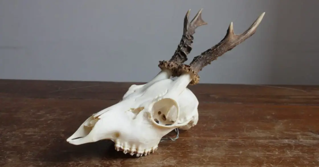
On this page we’ll share information about deer skull anatomy, including the parts of a deer’s skull. Whether you’re interested in a skull as a decor piece, or have interest for scientific research purposes, some of the information on this page may be useful.
(We will be updating the content on this page in the coming weeks as we continue to make broad updates across this website to ensure all information provided is accurate, relevant, and up to date.)
The Pudú (Pudu puda), one of the smallest deer skull in the world and indigenous to Chile, is considered vulnerable (VU) throughout the national territory, there is little specific information on the organic systems that compose it, and it is becoming more common find them in urban areas. These problems awaken the need to strengthen research and study of this species. Consequently, in the present study an anatomical description of the cranial skeleton of two specimens of Pudú puda was made, where their main bony characteristics were highlighted by comparing them with domestic species already studied (sheep and goats). Thus, it was possible to determine differences between the species studied and the need to deepen the analysis through measurements of the bone pieces.
INTRODUCTION
The Pudú is the smallest of the native deer of Chile (and in fact one of the smallest deer in the world). Within the scientific sector the species is also called Pudu puda. Its distribution occurs between Maule and Aysén, possibly in the north of Magellan and abundant on the island of Chiloé.
This is a species, with little specific information regarding the different organic systems that compose it, and its presence in urban areas is increasing, due to the increasing development of cities. This generates, the need to enhance knowledge about the morphology of this species.
The present study is the beginning of an extensive but necessary work that seeks to contribute to the development of animal anatomy and aims to make an anatomical description of the bone segments that make up the skull of two specimens (female and male) of the pudú species, bearing relation to the directional terms and planimeters expressed in the current veterinary anatomical payroll (International Committee on Veterinary Gross Anatomical Nomenclature, 2012) and schematically considering the two main regions of division, cranial or neurocranial region and facial or splenocranial region, as well as each subregion that compose them. Also separating in detail the visible structures on the external and internal surface of the cranial cavity.
The results of this study sought to found a solid base in the knowledge of bone architecture and thus contribute and as a fundamental piece in the development of morphology in wildlife species.
MATERIAL AND METHOD
The material associated with this study is biological. In which the skulls of two adult specimens (female and male) of the Pudu puda species were used, both obtained in years prior to this study through the osteotechnical technique in the Veterinary Anatomy unit of the Santo Tomás University, ( UST) headquarters Puerto Montt, Chile.
The anatomical description addresses the structures under study from an overview to a decomposition of such a structure, in order to guide the reader spatially and topographically as well as in their general shape and appearance, and then carefully characterize each of the segments and pieces that make up that whole.
Consequently, the study method that was used is of a qualitative descriptive type. In which the observation and description of bone pieces (skull) was performed.
Descriptive development followed a methodical order; where the concepts used to refer to the different structures (sight and planes of observation, topography, spatial arrangement, underlying relationships and names) were taken from the veterinary anatomical payroll (International Committee on Veterinary Gross Anatomical Nomenclature); beginning with a general view in the different planes of the cranial skeleton, to continue with a schematic division of the two main segments that make up the skull (neurocranium and esplacnocráneo) and each of the bones that make up these bone segments.
The results obtained from the anatomical description of the skull were analyzed and discussed under criteria of absence or presence of structures with respect to that described in the anatomical bibliography for small domestic ruminants (such as the Caprino or the Ovino). This is based on the fact that there is a taxonomic and phylogenic relationship between the Pudú and this group of species, categorizing them as at least small ruminants, as mentioned by Guzmán (2007 ) in his genetic study of the Pudú, where he describes that the Bovidae and Cervidae families show a certain degree of relationship.
The objective of this work is nothing more than to describe the cranial osteology of the Pudu puda species and the comparison with other small ruminants, is based on the fact of making the results even more conclusive and giving the reader a reference to the architecture bone of small domestic ruminants.
The bone pieces that were used in the development of this work belong to the veterinary anatomy unit of the Santo Tomás University headquarters in Puerto Montt and were obtained years prior to this study using the osteotechnical technique. This technique was carried out on preserved Pudú corpses, both obtained by donation from SAG Puerto Montt officials for educational purposes.
For in-depth observation of the different segments of the Pudú skull, a cut was made in the median sagittal plane of the cranial skeleton and a cut in the dorsal plane of the neurocranium. This allowed an appreciation of the skull architecture from both a general external view and an internal view of the nasal cavity and the endocranial cavity.
RESULTS AND DISCUSSION
Cranial Skeleton . The deer skull or cranial skeleton of the Pudu puda species, as in all vertebrates, is made up of two main segments, neurocranial and splenochronous. Already from the embryonic stage there is a clear differentiation between both future bone segments and in mammals in the adult state, it generally manifests itself with a greater and broad development of the facial bones (splanchnochronous) compared to the bones of the neural (neurocranial) part, similar to raised in ruminant anatomy publications.
When making a general appreciation of the deer skull, it is observed to be more stylized, long and homogeneous in relation to small domestic ruminants (goats and sheep), given this possibly due to the greater angle to which the neurocranium and the esplachnocranium are related, by the prolongation of the frontal and parietal bone in the formation of the neurocranial ceiling, as well as by the decrease in the dorso-ventral axis of the maxillary bones in the formation of the face. In addition, the smaller lateral projection of the margins of the eye socket, give this homogeneous and stylized aspect to the esplachnocranium, the latter possibly determining the most frontal focus of the characteristic eyeballs of this deer species.
Fig. 1 Rostral view of the deer skull of a male specimen of the Pudu puda species, showing the lateral limits of the esplachnocranium, given by the lateral margin of the zygomatic bone. Source: Veterinary Anatomy Unit, UST, Puerto Montt headquarters, Chile.
Fig. 2 Caudal view of the cranial skeleton of a male specimen of the Pudu puda species, showing the external occipital protuberance (A). Source: Veterinary Anatomy Unit, UST, Puerto Montt headquarters, Chile.
Fig. 3 Ventral view of the deer skull of a male specimen of the Pudu puda species, showing the muscular processes of the basilar portion of the occipital bone (A and A ”). Source: Veterinary Anatomy Unit, UST, Puerto Montt headquarters, Chile.
Neurocranium
Occipital bone . In the occipital bone of the Pudú three parts are observed, basilar, lateral and scaly. In the latter, the external occipital protuberance is observed, forming in its entirety the dorsal margin of the caudal wall of the cranial cavity, it stands out for being quite defined, it projects laterally where it articulates with the caudal margin of the temporal bone in its portion petrous internally and tympanic externally. While in the basilar portion, the muscular tubercles stand out, which are closer to the mid sagittal line, the rostral end of this portion being narrower and thus giving a triangular shape to the base of the neurocranium.
Sphenoid bone
Basisphenoids . Due to the proximity of the muscular tubercles to the median sagittal angle in the basilar portion of the occipital, the body of the basisfenoid maintains this congruence in its shape and also adopts a triangular and narrower shape, unlike what occurs in the goat and sheep, where the appearance is rectangular. In an endocranial view of the body of the basisfenoides, the pituitary fossa and the dorsum selate are seen together with their clinoid processes as the caudal limit.
Fig. 4 Ventral view of the cranial skeleton of a male specimen of the Pudu puda species, showing the basisfenoid bone. Source: Veterinary Anatomy Unit, UST, Puerto Montt headquarters, Chile.
Presphenoids . The presphenoid bone continues rostrally from the basisfenoid forming the base of the skull. In an internal view and after the pituitary fossa formed by the basisfenoid body, its wings are seen lifting the floor and thus narrowing the cranial cavity, to then articulate with the orbital portion of the frontal bone.
Fig. 5 Dorsal view of the endocranial cavity of a female specimen of the Pudu puda species in which the following stand out: the wings of the presphenoid (A), ethmoid bone (B). Source: Veterinary Anatomy Unit, UST, Puerto Montt headquarters, Chile.
Temporary bone . As in other small domestic ruminants, the temporal bone of the Pudú participates in the formation of the lateral walls of the cranial cavity. The observation highlights the loss of continuity between the lateral margins of the caudal wall of the cranial cavity and the birth of the zygomatic process from the squamous portion of the temporal, given this by the dorsal lateral projection of the external acoustic meatus. Situation different from what happened in sheep and goats, where the external acoustic meatus projects laterally ventrally at the beginning of the zygomatic process. Keeping similarity to what has been stated in publications on ruminant animals (Sisson & Grossman; Gloobe; Shively; Köning & Liebich).
Fig. 6 Left lateral view of the deer skull of a male specimen of the Pudu puda species in which the following stand out: Temporal bone (A), external acoustic meatus (B). Source: Veterinary Anatomy Unit, UST, Puerto Montt, Chile.
Parietal bone . In the Pudú, the parietal bone reaches a great extension, forming a large part of the roof and the lateral walls of the cranial cavity. The suture through which it articulates on its rostral margin with the frontal bone, marks the birth in caudal projection of the cornual processes of the frontal bone, a situation similar to that which occurs in the goat but very different in the sheep.
Frontal bone . It is an extensive bone that forms the rostral half of the roof of the cranial cavity, the other half is completed by the parietal bone. Laterally in the squamous portion, the zygomatic process that participates in the formation of the dorsal caudal margin of the ocular orbit is projected, which unlike other small domestic ruminants, this margin does not protrude from the lateral limits of the face, thus giving an appearance sharper to the deer skull. Here is also the supraorbital foramen.
The cornual processes, present only in the male, project in the caudal direction from the caudal margin of this bone. At the birth of the cornual processes, a depression or fossa stands out, which also coincides with the birth of the zygomatic process of the frontal bone.
The ethmoid bones, hyoid and interparietal apparatus, with similarities in their architecture in relation to small domestic ruminants.
Fig. 7 Dorsal view of the deer skull of a male specimen of the Pudu puda species showing: Parietal bone (A), Frontal bone (B), fossa at the origin of the cornual processes and zygomatic processes of the frontal bone ( C), Lateral margin of the face marked by the zygomatic bone (D), Supraorbital hole (E), Cornual processes (F). Source: Veterinary Anatomy Unit, UST, Puerto Montt headquarters, Chile.
Scalanochronous
Maxillary bone . The maxillary bone has an elongated shape on its face-caudal axis and irregular and narrow on its dorso-ventral axis, this is partially influenced by the presence of a large lacrimal bone and its wide external fossa, however, its rostral margin has a smaller angle when articulating with the incisor bone, which gives a less sharp appearance when compared to the spleno-skull of other small small ruminants.
On the external surface, a less developed and less prominent facial tubercle is observed than in other small ruminants, even located under the eye socket and zygomatic bone. While the infraorbital hole is in a rostral arrangement on the first premolar.
As for the denture, the upper arch shows: 6 pieces, 2 x (3 premolars and 3 molars); Without upper incisors.
————–
Fig. 8 Left lateral view of the deer skull of a male specimen of the Pudu puda species in which the following stand out: Incisor bone (A), Maxillary bone (B), Joint angle between H. palatine and H. maxilla, Infraorbital hole (D), Facial tubercle (E). Source: Veterinary Anatomy Unit, UST, Puerto Montt headquarters, Chile.
Incisor bone . The incisor bone presents a smaller angle with respect to the joint with the maxillary bone at its rostral end, unlike what occurs in other small domestic ruminants.
Nasal bone . As in small domestic ruminants, the nasal bone is an even bone and forms the roof of the nasal cavity. In Pudú it stands out for being pointed at its rostral end, presenting a nasoincisive notch and also for being divided into its major axis up to the Rostral half through a bone incision.
Fig. 9 Dorsal view of the deer skull of a male specimen of the Pudu puda species in which the following stand out: nasal bone (A), incision of the nasal bone (B). Source: Veterinary Anatomy Unit, UST, Puerto Montt headquarters, Chile.
Palatine bone . Regarding the formation of the hard palate and the articulation with the palatal process of the maxilla through the horizontal lamina, there are no differences between the Pudú and other small domestic ruminants (Sisson & Grossman; Gloobe; Ashdown & Done, 2011). However they stand out; the presence of a pair of processes located in the perpendicular lamina, at the birth of the nasopharyngeal meatus and choanae, and the smaller diameter of the perpendicular lamina and therefore the lesser depth of the nasopharyngeal meatus. The latter is possibly given by the position and angle of the neurocranium with respect to the splanchnocranium, where in a general view of the skull a greater angle is seen and therefore a more stylized cranial skeleton.
Lacrimal bone . The lacrimal bone has a great development and there are two surfaces (facial and orbital). On the orbital surface the lacrimal bulla is observed and below it the beginning of the nasolacrimal duct. While a large external lacrimal fossa is observed on the facial surface, it stands out for having great depth. Between the lacrimal, maxillary, nasal and frontal bone, a perforated opening is generated that communicates with the paranasal sinuses and nasal cavity, described only in the goat.
Zygomatic bone . It is presented articulated from its rostral end to the maxillary bone, with a caudal view where it articulates with the frontal and zygomatic bone. It stands out for lacking lateral projection, being thinner than flattened unlike what occurs in other small domestic ruminants.
Jawbone . The mandible is made up of two segments, right lateral and left lateral, each presenting a body and a branch (Sisson & Grossman; Gloobe; Shively). It stands out in the branch, a prominent and accentuated angular process, given by the presence of a marked notch in the limit with the body by the ventral margin, with greater development in males than in females. For its part, the coronoid process stands out for its great caudal projection, being also greater in males. Regarding the denture, the presence of 2x (4 incisors, 1 canine, 2 premolars, 3 molars) is observed (Figs. 11 and 12).
Fig. 10 Oblique side view of the cranial skeleton of a male specimen of the Pudu puda species, in which the following stand out: Opening between the frontal, nasal, maxillary and lacrimal bones, associated with the functionality of the lacrimal system (A), fossa Lacrimal lacrimal bone external (B), Zygomatic bone (C). Source: Veterinary Anatomy Unit, UST, Puerto Montt headquarters, Chile.
Fig. 11 Left lateral view of the jaw of a male specimen of the Pudu puda species in which the following stand out: The body of the jaw (A), branch of the jaw (B), cut-out of the mandibular angle (C), Process coronoid (D). Source: Veterinary Anatomy Unit, UST, Puerto Montt headquarters, Chile.
Fig. 12 Left lateral view of the mandible of a female specimen of the Pudu puda species in which the following stand out: The angular process and the notch of the angular process (A), coronoid process (B). Source: Veterinary Anatomy Unit, UST, Puerto Montt headquarters, Chile.
CONCLUSIONS
The observations made on the deer skull of the Pudu puda species and the subsequent description of the bony segments that compose it, determine that there are differences with respect to the skull of small domestic ruminants.
It was possible to emphasize the main differences existing at the level of the neurocranium and the esplano-skull, presenting extensions, depressions and pits that highlight the architecture of the skull of this species.
It is necessary to consider that the anatomical description is not enough to establish relationships or quantitative parameters at the level of the skull of this species, which is why the continuity of this study deepened in morphometric dimensioning, each observed difference.


